
Considering just how much we stand on them and what we ask of them, it’s surprising just how much we ignore our feet. Whether it is forcing them into ill-fitting or high-heeled shoes, or bearing the weight of us all day and everyday, our feet remain relatively uncared for. But healthy feet and ankles are essential for walking, running and jumping. No wonder they are sometimes called ‘little soldiers’.
The pressure on the foot increases according to the activity they are bearing, two-fold when walking and four-fold when running.
Foot Structure

The foot is made up of 26 bones. They are aligned in tiers, the hindfoot, midfoot and forefoot:
- Hindfoot: talus and calcaneus
- Midfoot: 5 tarsals which include the cuboid, navicular and cuneforms
- Forefoot: Metatarsals and phalanges
The foot is usually grouped with the ankle joint – or talocrural joint – made up of the tibia, fibula and talus. This joint allows plantar flexion and dorsiflexsion. The movement respectively of the foot away from and towards the shin bone.

The Foot’s Arches
The midfoot provides the shaping of the foot’s arches. The stresses on the foot means that there are actually 3 arches in the foot. The medial longitudinal arch running along the length of the inside of the foot that we are most aware of; the lateral longitudinal arch on the outside of the foot; and the transverse arch running along the balls of the feet. The arches raise the centre of the foot to distribute and absorb the body’s weight. The medial longitudinal arch is most prominent in this, but it is subject to collapse giving rise to the term ‘fallen arches’. The strength of the soft tissue – principally the adductor hallucis – is largely responsible for maintaining the structure of the arch so a collapse can result in the wearing of aids such as orthotics.
Muscles and soft tissue of the foot
There is a strong relationship between the muscles of the lower leg and foot because of their attachment points. The calf muscles gatrocnemius and soleus form the Achilles (Calcaneous) tendon which attaches to the foot’s calcaneus. The peroneals, extensor and flexor muscles at the side of the leg attach at the toes.
Muscles within the foot itself are multiple but the most prominant is the flexor digitorum brevis on the plantar side of the foot, giving it it’s fleshy underside. But collectively they can be referred to as the plantar tissues and plantar fascia.
Foot structural problems
The feet do not escape postural problems. One look at your shoes to see the wearing down of the heal on the lateral or medial side will reveal a tendancy to pronate or supinate the feet.
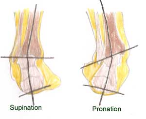
In supination, the foot is inverted and the body weight is carried on the lateral side of the foot. You can see that the calcaneal bone and the lateral ankle bone (malleoli) become distorted.
More common is foot pronation where the foot is everted and body weight is carried on the medial side of the foot. The knees are forced to turn inwards also causing a similar internal rotation of the hip.
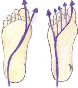
Over pronation can lead to other problems as poor muscle function. In normal foot planting when walking and running the foot is slightly supinated and weight is carried on the lateral side of the foot. Over-pronation means the medial longitudinal arch is flattening and less able to absorb shock and weight is distributed towards the big toe.
21 January 2016